Quick Takeaway
- Atrophic gastroenteritis thins the intestinal lining, causing villous loss.
- Reduced surface area impairs absorption of proteins, fats, carbs, and key vitamins.
- Typical deficiencies include iron, vitamin B12, calcium, and fat‑soluble vitamins.
- Diagnosis relies on endoscopic biopsy and targeted blood tests.
- Effective management combines diet changes, enzyme supplements, and probiotic therapy.
What Is Atrophic Gastroenteritis?
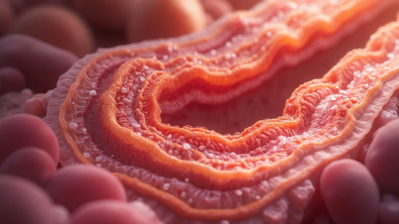
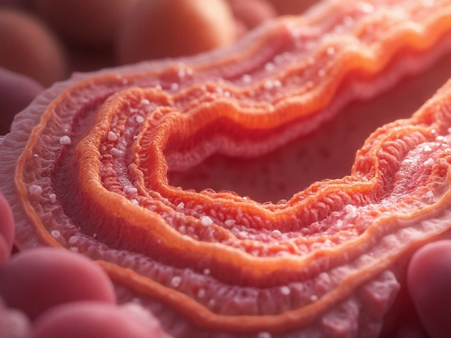
Atrophic gastroenteritis is a chronic inflammatory disorder of the small intestine that leads to progressive thinning (atrophy) of the mucosal lining. It is characterised by loss of villi, crypt hyperplasia, and chronic infiltration of inflammatory cells. The condition often follows long‑standing celiac disease, tropical sprue, or severe infections, but it can also arise idiopathically.
How the Small Intestine Structure Changes
The small intestine is the primary site for nutrient extraction, thanks to its finger‑like projections called villi. In atrophic gastroenteritis, villous atrophy reduces the absorptive surface by up to 80% in severe cases. Crypts, the glandular pits between villi, become deeper and more proliferative, further distorting the mucosal architecture. This structural breakdown directly limits the contact time between digested food and transport proteins, sabotaging the uptake of almost every nutrient.
Mechanisms Behind Impaired nutrient absorption
Three intertwined mechanisms explain the malabsorption:
- Surface loss: With fewer villi, the area available for carrier‑mediated transport shrinks dramatically.
- Enzyme deficiency: Brush‑border enzymes (e.g., lactase, maltase) are produced by enterocytes that disappear in atrophy, limiting carbohydrate breakdown.
- Transport protein down‑regulation: Inflammation suppresses expression of carriers for iron (DMT1), calcium (TRPV6), and B12‑intrinsic factor complex.
Because each nutrient follows a specific uptake route, the pattern of deficiencies can clue clinicians into which pathways are most compromised.
Clinical Consequences: Common Deficiencies
Patients with atrophic gastroenteritis often present with a cluster of micronutrient deficiencies:
- Iron‑deficiency anemia: Iron is absorbed mainly in the duodenum via DMT1; villous loss here drops serum ferritin quickly.
- Vitamin B12 deficiency: B12 requires intrinsic factor binding and uptake in the distal ileum. Atrophy disrupts both the binding surface and ileal transport.
- Calcium and Vitamin D insufficiency: Calcium uses active transport (TRPV6) and passive diffusion; both suffer when the mucosa thins, leading to osteopenia.
- Fat‑soluble vitamin (A, D, E, K) malabsorption: Lipid droplets need micelle formation and chylomicron assembly, both hindered by reduced enterocyte mass.
In severe cases, protein‑calorie malnutrition can develop, evident as weight loss, muscle wasting, and edema.
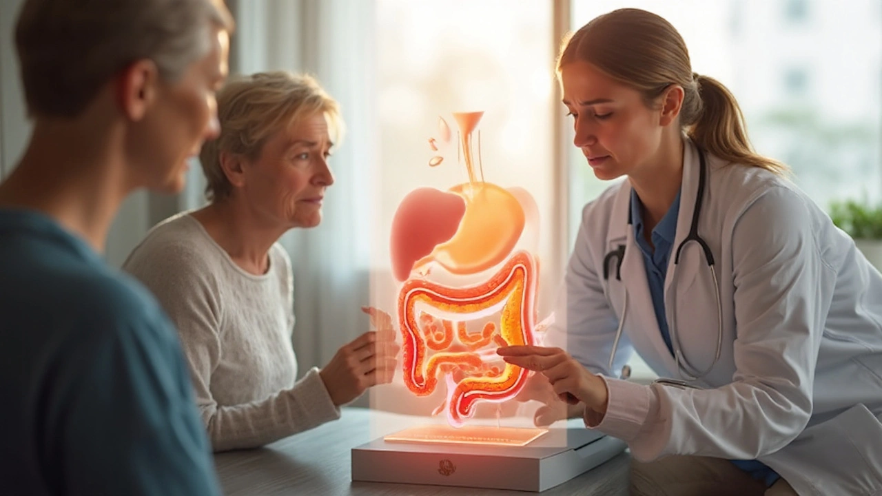
Diagnosis and Assessment
Accurate diagnosis hinges on visual and histologic evidence. An endoscopic biopsy of the duodenum or jejunum reveals flattened villi, crypt hyperplasia, and increased intra‑epithelial lymphocytes. Blood work complements the picture: low hemoglobin, low ferritin, low serum B12, and low 25‑OH vitamin D levels signal malabsorption. A stool fat analysis (72‑hour collection) can quantify fat malabsorption, useful when patients report steatorrhea.
Management Strategies
Tackling atrophic gastroenteritis requires a two‑pronged approach: heal the mucosa and replace lost nutrients.
1. Dietary Modifications
- Adopt a low‑residue, easily digestible diet during acute phases-think boiled vegetables, lean proteins, and rice.
- Once inflammation eases, introduce fermented foods (yogurt, kefir) to restore a healthy gut microbiome.
2. Probiotic therapy
Specific strains such as Lactobacillus rhamnosus GG and Bifidobacterium longum have shown in clinical trials to reduce intestinal inflammation and modestly increase villous height within 12 weeks.
3. Enzyme replacement
Pancreatic enzyme preparations (amylase, lipase, protease) help compensate for brush‑border loss, especially when patients report persistent bloating after fatty meals.
4. Targeted dietary supplementation
- Iron: ferrous sulfate 325mg twice daily, preferably with vitamin C to boost absorption.
- Vitamin B12: 1000µg oral cyanocobalamin weekly, or monthly intramuscular injections for severe cases.
- Calcium + Vitamin D: calcium carbonate 500mg with vitamin D3 800IU twice daily.
- Fat‑soluble vitamins: water‑soluble formulations (e.g., vitamin E 400IU daily) improve bioavailability.
Regular monitoring every 3-6 months ensures that levels are rising and that excess supplementation does not cause toxicity.
Comparison with Similar Gastrointestinal Conditions
| Condition | Primary Pathology | Typical Deficiencies | Diagnostic Hallmark | First‑Line Treatment |
|---|---|---|---|---|
| Atrophic Gastroenteritis | Villous atrophy + chronic inflammation | Iron, B12, Ca, fat‑soluble vitamins | Blunted villi on biopsy | Diet, probiotics, enzyme & nutrient supplements |
| Celiac Disease | Autoimmune reaction to gluten | Iron, folate, B12, D | Positive anti‑tTG antibodies + villous flattening | Strict gluten‑free diet |
| Crohn’s Disease (small‑bowel) | Transmural inflammation, strictures | Vitamin B12, iron, fat‑soluble vitamins | Skip lesions on endoscopy | Immunosuppressants, biologics |
| Normal Mucosa | Intact villi, no inflammation | None (full absorption) | Healthy villous height on biopsy | Not applicable |
Notice how atrophic gastroenteritis shares the malabsorption pattern of celiac disease but lacks a clear dietary trigger. This nuance guides clinicians toward broader anti‑inflammatory and microbiome‑focused therapies.
Related Concepts and Next Steps
Understanding atrophic gastroenteritis opens doors to several adjacent topics within the digestive‑health cluster:
- Intestinal permeability ("leaky gut"): Chronic inflammation often increases tight‑junction disruption, exacerbating systemic inflammation.
- Small‑intestine bacterial overgrowth (SIBO): Stagnant luminal content can foster bacterial proliferation, further impairing absorption.
- Autoimmune enteropathy: An even rarer cause of villous loss that requires immunosuppression.
Readers who want to dive deeper might explore "How to Diagnose SIBO" or "Nutrient‑dense Foods for Malabsorption" as natural follow‑up articles.
Frequently Asked Questions
Can atrophic gastroenteritis be reversed?
Yes, in many cases. Removing the inflammatory trigger, supporting the gut microbiome, and providing targeted nutrient supplements can stimulate villous regrowth within 6-12 months. Early intervention yields the best outcomes.
What tests are most reliable for detecting malabsorption?
A combination of upper‑GI endoscopy with duodenal biopsy, serum micronutrient panels (iron, B12, 25‑OH vitamin D), and a 72‑hour fecal fat test provides a comprehensive picture. Breath tests for SIBO can also be informative.
Is a gluten‑free diet enough to treat atrophic gastroenteritis?
Only if gluten is the underlying trigger (e.g., celiac disease). In pure atrophic gastroenteritis without gluten sensitivity, the diet alone rarely restores villous architecture; probiotics, enzymes, and supplements are essential.
How long should I take iron supplements after diagnosis?
Typically 3-6 months, or until ferritin normalises and hemoglobin stabilises. Continued monitoring prevents overload, especially if the gut heals and absorption improves.
Can probiotics worsen symptoms?
Rarely, but in heavily inflamed mucosa some patients experience gas and bloating. Start with low‑dose, single‑strain products and increase gradually while tracking symptoms.
What lifestyle changes help protect the gut?
Avoid smoking, limit NSAID use, manage stress (mindfulness, light exercise), and maintain a diet rich in soluble fiber and fermented foods. These habits support mucosal healing and microbiome balance.


charlise webster
Actually, the villous atrophy isn’t just a side effect; it’s the primary driver of the malabsorption you described.