Intestinal Villous Atrophy: What You Need to Know
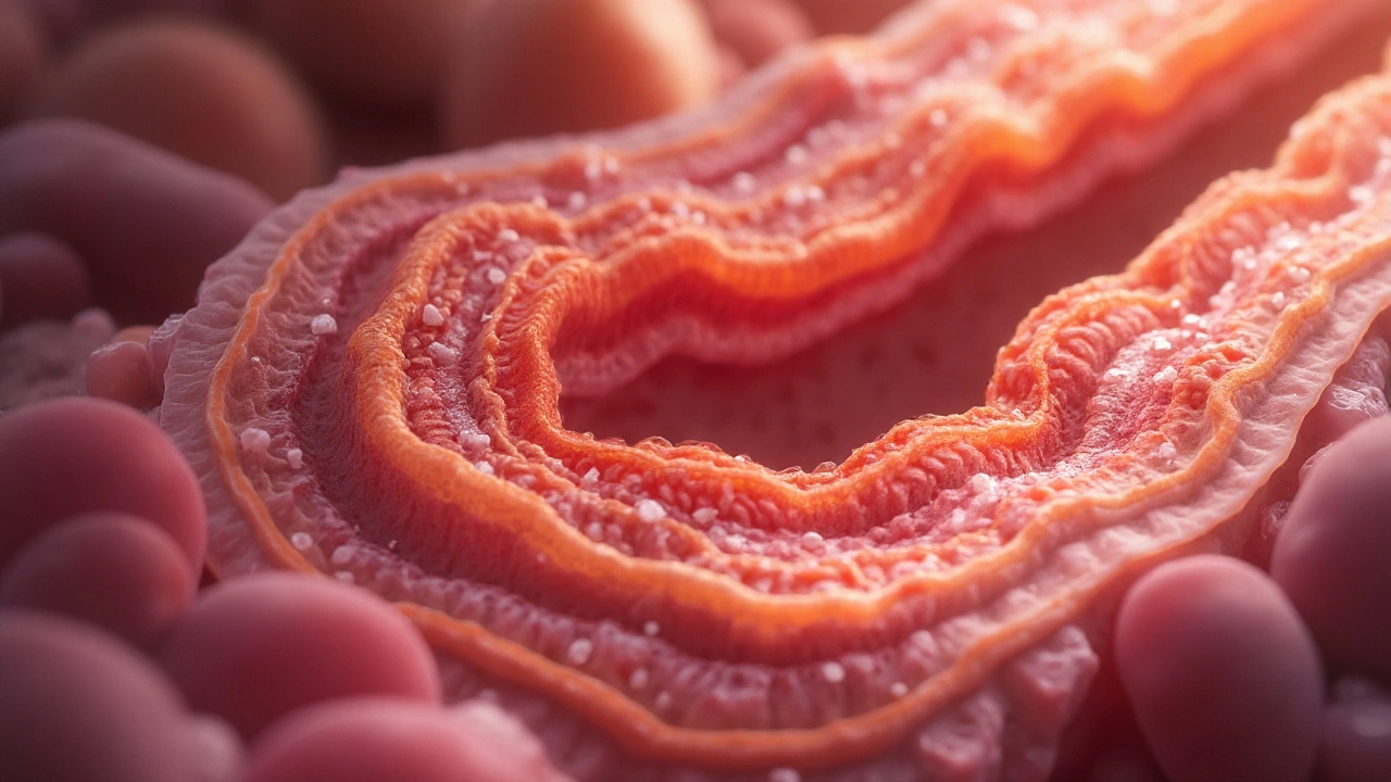
When dealing with intestinal villous atrophy, the flattening of the tiny finger‑like projections (villi) that line the small intestine, reducing nutrient absorption. It’s most commonly linked to celiac disease, an autoimmune disorder triggered by gluten that attacks the intestinal lining, which in turn creates malabsorption, the inability to absorb vitamins, minerals, and calories properly. The condition often forces patients onto a strict gluten‑free diet, a lifelong eating plan that removes wheat, barley, and rye to allow the gut to heal and may require supplementation to correct nutrient deficiencies, shortages of iron, calcium, vitamin D, and B‑vitamins caused by poor absorption. Understanding intestinal villous atrophy helps you see why each piece matters and sets the stage for effective care.
Why the Villi Flatten: Triggers and Mechanisms
The core triple here is simple: gluten exposure + genetic susceptibility = autoimmune attack → villous atrophy. People who carry the HLA‑DQ2 or HLA‑DQ8 genes react to gluten peptides, prompting immune cells to release inflammatory cytokines that erode the villi. This process not only flattens the surface but also raises intestinal permeability, sometimes called “leaky gut,” which can let undigested particles trigger further immune responses. In short, the disease demands both a trigger (gluten) and a predisposition (genetics) to damage the gut lining.
Symptoms usually reflect the loss of surface area. Common complaints include chronic diarrhea, bloating, weight loss, and fatigue. Children may experience growth delays, while adults often report iron‑deficiency anemia or bone pain. Because the signs overlap with other GI disorders, doctors rely on specific tests to confirm the diagnosis.
Diagnosis hinges on three steps: serology, imaging, and biopsy. Blood tests look for anti‑tTG (tissue transglutaminase) and EMA (endomysial) antibodies, which are elevated in most cases. An upper‑endoscopy lets doctors take a tiny piece of the duodenum to examine under a microscope. The hallmark finding is flattened villi with increased intra‑epithelial lymphocytes. When the biopsy shows these changes and serology is positive, the diagnosis of celiac‑related intestinal villous atrophy is solid.
Treatment is straightforward but lifelong. The first and most critical step is eliminating gluten from the diet, which usually restores villous architecture within months to a couple of years. Nutritionists often recommend a high‑fiber, naturally gluten‑free diet rich in fruits, vegetables, lean proteins, and gluten‑free whole grains like quinoa or buckwheat. Because absorption may still be impaired early on, doctors frequently prescribe iron, calcium, vitamin D, and B‑vitamin supplements to bridge gaps until the gut heals.
If left unchecked, persistent villous atrophy can lead to serious complications: osteoporosis from calcium loss, refractory anemia, infertility, or even an increased risk of intestinal lymphoma. Regular follow‑up appointments with blood work and occasional repeat biopsies help ensure the gut stays on track. Lifestyle tweaks—reducing alcohol, avoiding NSAIDs, and managing stress—also support healing, as inflammation can worsen the condition.
Below you’ll find a curated set of articles that dive deeper into related topics such as how stress influences clotting, the impact of environmental toxins on hormonal health, and the role of probiotics in gut disorders. Each piece adds a layer to the big picture of gut health, medication safety, and lifestyle choices that can affect intestinal villous atrophy and its management.
Explore how atrophic gastroenteritis damages the gut lining, blocks nutrient uptake, and leads to specific deficiencies, plus diagnosis and treatment tips.
Continue reading...